cranial nerves are directly attached to the brain, they can be sensory, motor or mixed nerves. we use roman numerals to name the nerves from I to XII. they send electrical signals between your brain and different parts of your face, neck, head and torso. these signals help us see, smell, taste, hear and move our facial muscles. the cranial nerves start at the back of our brain and play a key part in our nervous system. we have 12 cranial nerve pairs, each pair splits to serve the two sides of our brain and body.
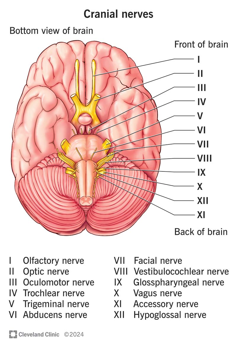
CN I - olfactory nerve: sense of smell. we have one pair of this nerve, one is on the left side of our brain and the other is on the right side. it is the shortest sensory nerve, it starts in our brain and ends in the upper inside part of our nose. this nerve is also apart of the autonomic nervous system (ANS) which regulates body functions. it detects odors, scents and aromas. substances that have smell give off tiny molecules that we inhale through our nose, special cells called olfactory receptors detect these molecules and relay the information to the brain through the olfactory nerve and allows us to perceive smell. the olfactory system turns on our sense of smell in two ways; first is nostrils as mentioned before and the back of the throat, drinking or chewing also releases those molecules, they travel up the throat to the olfactory receptors in the back of the nose. olfactory mucosa also plays an important role, it is located in the upper part of the nasal cavity and contains 3 different types of cells: olfactory receptor cells that support two processes: dendritic process which propels cells towards tiny hairs in the olfactory mucosa where they stimulate olfactory cells and central process which directs cells in the opposite direction. sustentacular cells which provide support to nearby tissue and basal cells which both olfactory receptor cells and sustentacular cells can develop from. the olfactory nerve is one of two nerves (visual nerve/CN II) that originate directly from the cerebrum which is the upper part of the brain. olfactory nerve fibers travel to an area in the upper part of the nose called the olfactory bulb but before it does the nerve passes through the cribriform plate which is a spony, lightweight skull bone that separates the nasal area from our brain.

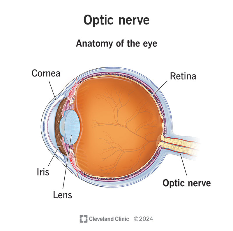
CN II - optic nerve: vision. the optic nerve is the connection that lets the eyes send signals to the brain describing what they see, the brain takes those signals and processes them and then uses them to construct what we see. this nerve also contributes to certain eye reflexes and our circadian rhythm which is our body's internal clock. it is comprised of millions of nerve fibers that send visual messages to the brain to help us see. we have an optic nerve at the back of each eye that directly connects to our brain. each nerve is a one-way connection and it only carries signals from the eyes to the brain. the retinas at the back of each eye detects light and converts that into electrical signals. the optic nerve carries those signals to the brain. this nerve is extra special because of how it forms, it's the only cranial nerve that is also part of the central nervous system (CNS). to get to the brain the optic nerves extend out from the retina and travel a route that includes the optic canal which is a bony opening that your optic nerves pass through to enter the skull and reach the brain. then it goes through the optic chiasm which is a y-shaped junction where nerve fibers meet up to travel together, some of the nerve fibers from both optic nerves switch sides. that crossover is a part of how the brain organizes left and right sided input from both eyes so it can merge them into the single and seamless pictures we see. the brain is just after the separate optic nerves that join at the optic chiasm. once in the brain they head straight to the visual cortex which is apart of the occipital lobe at the back of the brain which is where most visual processing happens. as the nerve fibers travel through the brain a small fraction of them branch off into other places in the brain. they support abilites like pupil reflexes, our pupils automatically adjust to let more or less light in, the reflex needs to be fast which is why it involves nerve fibers that branch off before they reach the visual cortex. next is the accommodation reflex which involves muscles in the ciliary body of each eye, those muscles adjust the shape of each eye's lens and thats how the eyes automatically focus on whatever we are looking at. last is the circadian rhythm which manages our natural sleep/wake cycle and contributes to many processes like body temperature, blood pressure and blood sugar. the optic nerve fibers tell the brain about the light they detected. most brains use that to help anchor the circadian rhythms to daytime and nighttime.
CN III - oculomotor nerve: eye muscle command signals. it's branches connect to muscles on multiple sides of the eyeballs which deliver movement commands from the brain to those muscles. this nerve lets us open our eyes, move our eyeballs up and down, inward and upward. it lets our eyes focus at different distances using the ciliary muscle to adjust lens shape. it also adjusts the pupils width to let more or less light into the eye. this cranial nerve works with many other systems including the vestibulo-ocular reflex which is combined with the vestibular system which controls balance with eye muscle and head movements to keep our gaze steady. it comes from 2 nuclei in the midbrain: oculomotor nucleus and the accessory parasympathetic nucleus or the Edinger-Westphal nucleus. this nerve has somatic and autonomic fibers. the somatic (voluntary) are bundled deep inside the nerve while autonomic (involuntary) fibers surround the somatic fibers around outside the nerve.
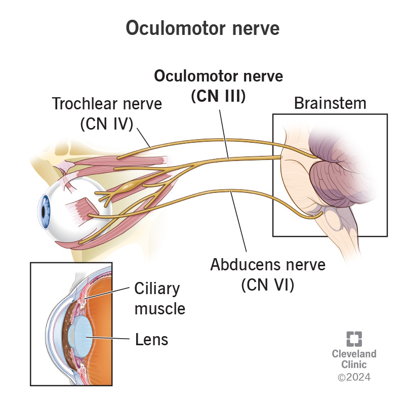
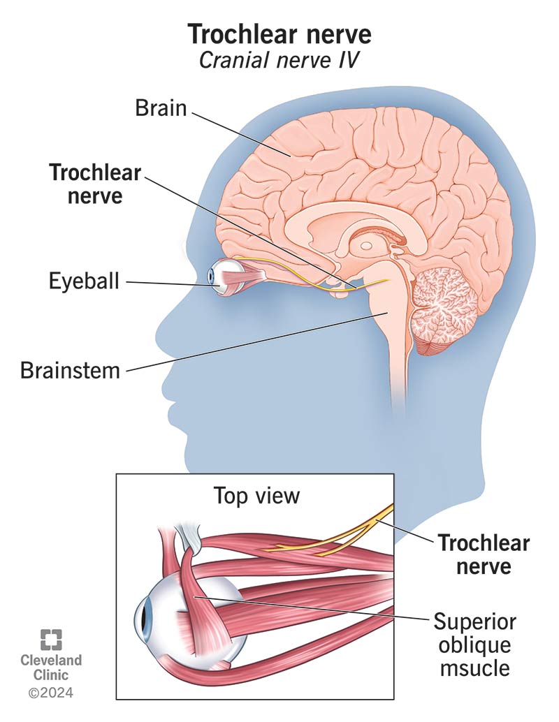
CN IV - trochlear nerve: we have two trochlear nerves one for each eye. this nerve has one job, to supply movement information to the superior oblique muscle. the trochlear nerve and superior oblique muscle makes it possible to look down they also enable us to move our eyes towards or aways from our nose. this nerve gets its name from "trochleae" which means pulley. this nerve controls the muscle from above which is connected to the eye near the top, the muscle goes through a sling of connective tissue that acts like a trochleae. it starts at the brainstem and passes through four areas before reaching the superior oblique muscle. the first area is the trochlear nucleus which is the closest to the brain. next is ambient cistern its near the brains outer protective tissue (the dura mater which is apart of meninges), third is the cavernous sinus which is a hollow space inside the skull. last is the orbit which is a bony socket of the skull that houses the eyeball, once it goes through the orbit the nerve attaches to the superior oblique muscle.
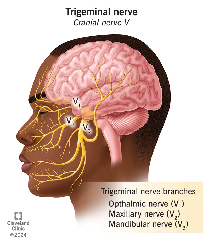
CN V - trigeminal nerve: this nerve helps the face recognize pain and touch, as well as temperatures. it also helps us chew. this is a 3 part nerve including opthalmic nerve (V1), maxillary nerve (V2) and mandibular nerve (V3). it sends signals from the brain to parts of the face and vice versa. we have two trigeminal nerves, one on each side of the face. the motor nerve fibers tell the muscles how and when to move, the sensory nerve fibers send pain, temperature and touch sensations from the skin to the brain. V1 nerve provides sympathetic nerve fibers which play a role in dilating the pupils and supplies sensation to the ciliary body, iris, lacrimal gland, conjunctiva and cornea. the V2 nerve relates to the upper jaw, it provides sensory information for portions of the nasal cavity, sinuses, maxillary teeth, palate and the middle portion of the face and skull specfically the area below the eyes and above the mouth. last is V3 which relates to the lower jaw, its the largest of the 3 and has both motor and sensory fibers. it provides sensory innervation of the buccal mucosa, floor of the mouth, mandibular teeth, tongue and the skin below the mouth. the motor part provides movement to all the muscles involved in mastication (chewing) these are the masseter, temporalis and pterygoid muscles. this nerve also plays a role in swallowing by supporting the digastric and myloyoid muscle. as said before we have two trigeminal nerves, one on each side of the head. they start in the brain and travel throughout the head and branch into 3. it begins within four nuclei in the brain. three of these are responsible for sensory and the last is for motor function. the 3 sensory nuclei merge to become one sensory root near the pons. this root becomes the trigeminal ganglion as it leaves the brainstem on each side. they are near the temples and is located within Meckels cave which is a space filled with cerebrospinal fluid between two layers of dura mater over the temporal bone. lastly the trigeminal ganglion splits into three trigeminal nerve branches aka ophthalmic, maxillary and mandibular branches. these branch out across each side of the head.
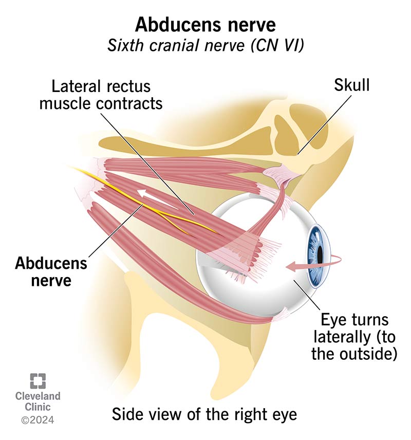
CN VI - abducens nerve: it controls the movement of the lateral rectus muscle of the eye. the muscle moves the eyes to the outside. moves left eye to the left and right eye to the right. it is a type of extraocular nerve which is a nerve outside the eye. it helps the brain communicate with your muscles. it works with the oculomotor nerve and trochlear nerve to move the eye muscles. it only has motor functions. it also works with the medical rectus muscle to make both eyes move to the left and right at the same time. it is a very small nerve behind the eyes. we have two of these nerves on the left and right side of the eyes. they extend from the brainstem and attaches to a muscle inside the eye socket, it passes through the subarachnoid space, dura mater and a canal through the skull into the eye socket where it meets the lateral rectus muscle. the abducens nucleus tells the nerve what to do, the nucleus is located in the pons which are in the upper back portion of the brain, they sit below the midbrain and above medulla oblongata which is above the spinal cord.
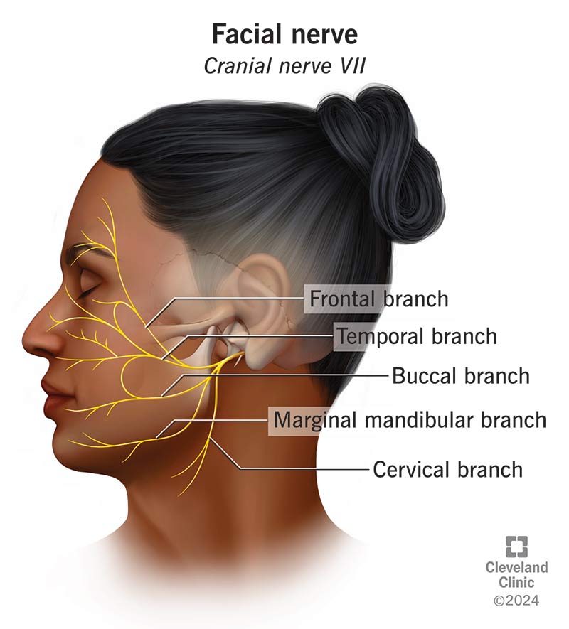
CN VII - facial nerve: a nerve in our head that sends signals from the brain to parts of our face and vice versa. we have two facial nerves, one on each side of the head. it contains motor, sensory and parasympathetic nerve fibers. the parasympathetic nerve fibers control the salivary glands and the lacrimal glands in the corners of the eyes, they trigger these glands to release saliva and tears. the sensory nerve fibers play an important role in hearing, they provide innervation to the ear canal, eardrum and outer ear. they also carry sensations from the front two-thirds of the tongue and lastly the motor nerve fibers have 5 branches: frontal (temporal) branches which control the forehead muscles, zygomatic branch which helps close the eyes, buccal branch which allows movement of the nose and blinking as well as raise the upper lip and corners of the mouth to form a smile. the marginal mandibular branches draws the lower lip down to form a frown, it travels through the middle ear to stapedius muscle which helps the inner ear respond to loud noises. last the cervical branch allows movement in the chin and lower corners of the mouth by controlling the platysma muscle in the neck. this nerve starts in the brainstem then travels through the base of the skull, it enters your face through an opening near the base of the ear, it then branches out through an opening near the parotid gland. which is a major salivary gland. from there the motor branches spread to parts of the face and neck.
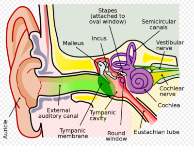
CN VIII - vestibulocochlear nerve: is a 2 part nerve that sends sensory information from the inner ear to the brain. this information is from the organs in the inner ear that helps us hear and maintain our balance. the first part is the vestibular nerve (vestibulo) that sends information from the vestibular system organs to the brain, those organs detect information about how our head is positioned and when it moves, the brain uses this information to help us stay balanced. those organs include two otolith organs which are the utricle and saccule then there are three semicircular canals, all five organs are filled with fluid lined with sensory cells that shift in response to our head movements. the signal is activated when the head moves, that fluid shifts which moves the sensory cells, that shift creates a signal about the bodys position and movement. neurons carry that signal along the vestibular nerve until it arrives at the vestibular nuclei complex in the brainstem which contains four nuclei, each nucleus in the brain is responsible for directing the signal to different parts of the body so we can maintain balance. some nuclei send sensory signals about the bodys position to the cerebral cortex and cerebellum, other nuclei send signals to the neck or legs to help change our position, others send signals to the eye muscles to help them stay focused even when we are moving that is called the vestibulo-ocular reflex or VOR. the second part is the cochlear nerve that sends audio information from the cochlea to the brain, the brain then uses this information to help us hear. the cochlea is also fluid filled and lined with sensory hair cells, the cells and fluid shift in response to sound waves. when sound waves reach the cochlea they vibrate the Basilar membrane, those vibrations cause sensory hair cells on that membrane to shift and that shifting creates a signal about sound quality, that includes the frequency of the sound waves (high/low the pitch is) and how loud. neurons carry that signal along the auditory nerve until it reaches the cochlear nuclei in the brainstem, the cochlear nuclei has 3 divisions, each one receives different information about sound, some receive information about low frequencies and others receive information about higher ones, the brain then processes that information the part that actually processes it is the auditory cortex in the temporal lobe. they join to form one nerve but still maintains the separate roles. this nerve passes through the internal auditory canal or IAC, this connects to the inner ear to the lower part of the skull. this nerve extends from the vestibular (Scarpas) ganglion which is located near the vestibular organs, it joins with the cochlear nerve inside the IAC to form this nerve. the vestibular nerve separates from the cochlear nerve once it reaches the vestibular nuclei complex in the brainstem, the cochlear nerve extends from spiral ganglion which is near the cochlea, it joins with the vestibular nerve inside the IAC to form this nerve, it then separates once it reaches the corresponding nuclei in the brainstem.
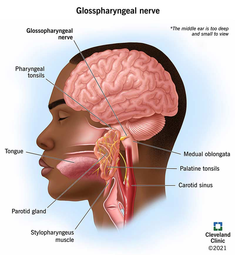
CN IX - glossopharyngeal nerve: this nerve is another CN with three types of fibers; motor, parasympathetic and sensory. it supports muscles, organs and body processes near the throat, includes the carotid sinus is a hollow area that helps drain blood from the brain into the carotid artery in the neck, it helps regulate blood pressure. next is the middle ear which has sensory nerve fibers that let us feel fullness from fluid buildup in the ears it also lets you feel an ear infection. the parotid (salivary) glands descrease saliva production when you finish eating. the stylopharyngeus muscle is a long muscle that runs down part of the throat, the nerve connects this muscle to lift the larynx (voice box) and pharynx and this lets us swallow safely. this nerve helps us taste food in the back third of the tongue and last the tonsils they are sensory nerve fibers that allow us to feel pain if we have a sore throat or swelling. the nerve starts at the lower part of the brainstem, it runs through the neck before reaching the throat it follows this path: first it exits the skull through a small opening called jugular foramen then it travels down the neck alongside the jugular vein, next it goes behind the styloid process which is apointy bone in the skull below the ear then it curves toward and touches the stylopharyngeus which is a muscle near the throat then last it passes under the hyoglossus muscle near the tongue.
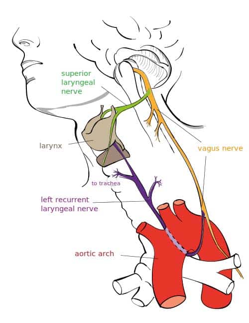
CN X - vagus nerve: also known as vagal nerves are the main nerves of the parasympathetic nervous system, it controls specific body functions such as digestion, heart rate and immune system. those functions are involuntary. they also play a role in breathing, mood, mucus and salvia production, skin and muscle sensations, taste, speech and urine output. this nerve is the longest CN running from the brain to the large intestine, the left vagus nerve goes down the left side and the right vagus goes down the right side of the body, "vagus" is latin for wandering. the nerve take a long course through the body, it exits the medulla oblongata in the lower brainstem and passes through/connect with the neck specifcally between the carotid artery and jugular vein, the thorax (chest), lungs, heart, abdomen and digestive tract. the left and right vagal nerves join to form the vagal trunk, they connect at the esophageal hiatus where the esophagus passes into the abdominal cavity, the trunk includes anterior and posterior gastric nerves that go to the abdomen. the vagal nerve branches include the inferior ganglion branch that serves muscles and nerves to your pharynx and larynx, the superior ganglion branch serves nerves to the ear and spine last is the vagus nerve branch that serves nerves to the lungs, heart and esophagus.
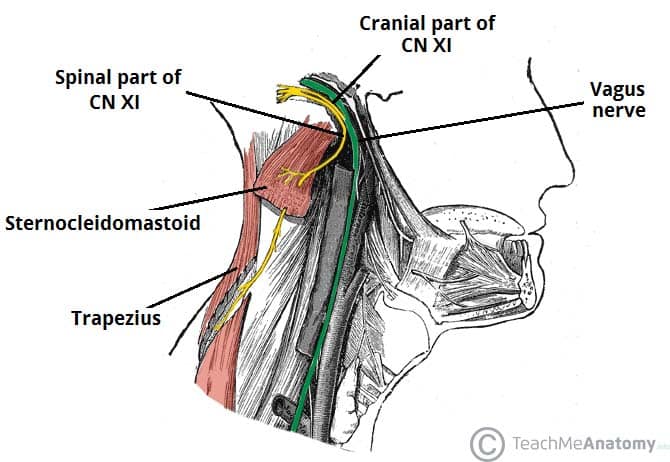
CN XI - spinal acessory nerve (SAN) is a motor nerve responsible for controlling the trapezius muscle shrugs the shoulders, assists in moving the head back and moves the scapula and sternocleidomastoid muscles (SCM) that rotates the head to opposite side and tilts the head to that side. unlike other CN this one comes from the spinal cord adn moves into the cranium trhough the foramen magnum then exists through the jugular foramen to innervate neck muscles. it arises from the nucleus ambiguss and connects with the vagus nerve (CN X) to give motor fibers to the pharynx, larynx and soft palate.
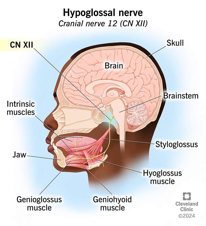
CN XII - hypoglossal nerve: starts at the base of the brain and travels down the neck and branches out ending at the underside and base of the tongue. the word "hypo" comes from greek which means under and "glossal" means tongue. this is a motor nerve that carries signals to and from the brain to control muscle movement. it controls muscles in the tongue allowing us to make noises with our mouth, swallow, speak and move food around in the mouth. this nerve controls the genioglossus muscles which push the tongue forward, the hyoglossus muscle that pulls the tongue back and flattens it, the intrinsic muscles change the tongues shape like curving and narrowing, last the styloglossus muscle which moves the tongue up and down. before it reaches the tongue the nerve travels down the spinal cord to the cervical plexus which is a complex nerve network that lets us feel sensation and movement in the trunk and neck, then it runs down the neck past the carotid artery and jugular vein then extends past the back of the throat until it reaches the floor of the mouth then branches off to connect with the different muscles.